
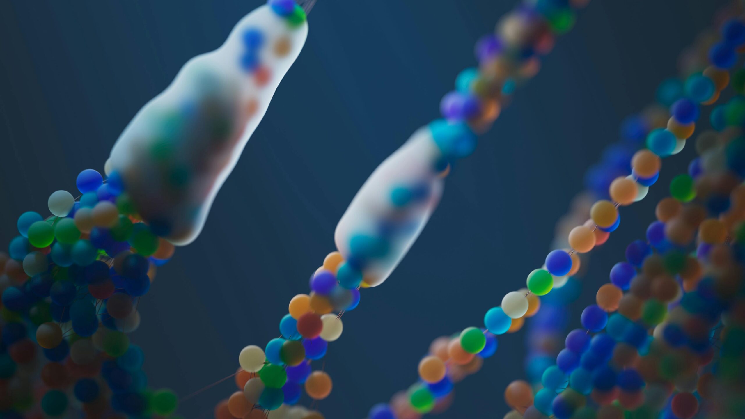
Modern science is unlocking groundbreaking insights into cellular behavior by comparing two-dimensional and three-dimensional structural matching techniques that revolutionize our understanding of biological systems.
🔬 The Revolution in Cellular Analysis Methods
The study of cellular structures has undergone a remarkable transformation over the past decades. Scientists have moved from simple microscope observations to sophisticated imaging techniques that reveal the intricate details of life at the molecular level. This evolution has brought forward an essential question: how do two-dimensional representations compare with three-dimensional models when analyzing cellular components?
Understanding this distinction is crucial for researchers, medical professionals, and biotechnology experts who rely on accurate cellular mapping to develop treatments, understand diseases, and advance scientific knowledge. The comparison between 2D and 3D matching methodologies represents more than just a technical debate—it’s a fundamental shift in how we perceive and interact with the building blocks of life.
Understanding Two-Dimensional Cellular Matching
Two-dimensional cellular matching has been the cornerstone of biological research for generations. This traditional approach involves examining cells and their structures on flat surfaces, typically through microscopy slides or digital imaging platforms. The technique offers several distinct advantages that have made it indispensable in laboratories worldwide.
The Strengths of 2D Analysis 💪
The primary advantage of two-dimensional matching lies in its accessibility and simplicity. Researchers can quickly prepare samples, capture images, and analyze patterns without requiring expensive equipment or complex computational resources. This efficiency makes 2D analysis ideal for high-throughput screening, where thousands of samples need examination within short timeframes.
Furthermore, 2D imaging produces clear, easily interpretable results. The flat representation eliminates depth-related complications, allowing scientists to focus on specific cellular features like membrane patterns, organelle distribution, or protein localization. Educational institutions particularly benefit from this approach, as students can grasp fundamental concepts without overwhelming complexity.
Limitations That Cannot Be Ignored ⚠️
Despite its utility, two-dimensional matching presents significant constraints. The most obvious limitation is the loss of spatial information. Cells exist in three-dimensional space, and flattening them onto a two-dimensional plane inevitably sacrifices crucial data about their true structure, volume, and spatial relationships with neighboring cells.
This dimensional reduction can lead to misinterpretations. For example, two structures that appear separate in 2D imaging might actually be connected in three-dimensional space. Similarly, the true size and shape of cellular components can be distorted when projected onto a flat surface, potentially leading to incorrect conclusions about their function or significance.
The Three-Dimensional Paradigm Shift
Three-dimensional cellular matching represents the cutting edge of biological imaging and analysis. This approach captures the complete spatial architecture of cells, providing unprecedented insights into their structure and function. Advanced technologies like confocal microscopy, electron tomography, and light-sheet fluorescence microscopy have made 3D reconstruction increasingly accessible.
Revolutionary Capabilities of 3D Matching 🚀
The power of three-dimensional analysis lies in its ability to preserve and reveal spatial relationships that are invisible in 2D representations. Researchers can now track cellular structures through their entire volume, understanding how organelles interact, how membranes fold, and how molecular complexes organize themselves within the cellular environment.
This comprehensive view has transformed numerous fields. In neuroscience, 3D matching enables scientists to trace complete neural circuits and understand synaptic connections. Cancer researchers can examine tumor microenvironments in their full complexity, identifying therapeutic targets that would remain hidden in flat images. Developmental biologists can watch tissues form and organs develop with unprecedented clarity.
Technical Challenges and Resource Requirements
However, three-dimensional cellular matching demands significantly more resources than its 2D counterpart. The equipment required for 3D imaging is expensive, often costing hundreds of thousands of dollars. Sample preparation becomes more complex, requiring specialized protocols to maintain cellular integrity throughout the imaging process.
Data management presents another substantial challenge. A single 3D cellular dataset can contain gigabytes or even terabytes of information, requiring powerful computers and sophisticated software for processing and analysis. The time investment increases dramatically, with some 3D reconstructions taking days or weeks to complete, compared to minutes or hours for 2D imaging.
Comparative Analysis: When to Use Each Approach
The choice between two-dimensional and three-dimensional cellular matching isn’t always straightforward. Both methodologies have specific applications where they excel, and understanding these contexts helps researchers select the most appropriate tool for their investigations.
Ideal Scenarios for 2D Matching 📊
Two-dimensional approaches remain optimal for several research contexts. Initial screening studies benefit from the speed and efficiency of 2D imaging, allowing researchers to identify promising candidates for further investigation. Quality control in pharmaceutical manufacturing often relies on 2D techniques to rapidly assess cell culture health and consistency.
Educational settings also favor 2D matching due to its lower cost and simpler interpretation. Students learning cellular biology can develop fundamental understanding without the complexity of three-dimensional visualization. Clinical diagnostics frequently employ 2D methods, where established protocols and rapid turnaround times are essential for patient care.
When 3D Becomes Essential 🎯
Three-dimensional matching becomes indispensable when spatial relationships are critical to understanding biological phenomena. Structural biology relies heavily on 3D reconstruction to determine protein conformations and molecular interactions. Tissue engineering requires precise three-dimensional mapping to design scaffolds that properly support cell growth and organization.
Drug development increasingly employs 3D cellular models to better predict how compounds will behave in living organisms. Traditional 2D cell cultures often fail to replicate the complex environment of tissues, leading to promising drugs failing in clinical trials. Three-dimensional matching in organoid cultures or tissue explants provides more reliable predictions of therapeutic efficacy.
The Technology Behind Modern Cellular Matching
Understanding the technological foundations of both 2D and 3D cellular matching illuminates their respective capabilities and limitations. Recent innovations have dramatically expanded what’s possible in both domains, though 3D technologies have seen particularly rapid advancement.
Imaging Modalities and Their Evolution 🔭
Traditional light microscopy remains the workhorse of 2D cellular imaging, supplemented by fluorescence techniques that highlight specific cellular components. Digital cameras and image processing algorithms have enhanced resolution and contrast, extracting maximum information from two-dimensional samples.
Three-dimensional imaging employs more sophisticated approaches. Confocal microscopy optically sections samples, capturing multiple focal planes that can be reconstructed into 3D volumes. Two-photon microscopy penetrates deeper into tissues with reduced photodamage. Electron microscopy achieves nanometer resolution in three dimensions through serial sectioning or tomographic reconstruction.
Computational Tools and Artificial Intelligence
The computational aspect of cellular matching has become increasingly important, particularly for 3D analysis. Machine learning algorithms now automatically identify cellular structures, track individual cells through time-lapse sequences, and quantify complex morphological features that would be impossible to measure manually.
Artificial intelligence has revolutionized pattern recognition in both 2D and 3D datasets. Neural networks trained on thousands of cellular images can detect subtle abnormalities, classify cell types, and even predict cellular behavior based on structural features. These tools democratize advanced analysis, making sophisticated techniques accessible to researchers without specialized computational expertise.
Real-World Applications Transforming Medicine and Research
The practical impact of comparing 2D versus 3D cellular matching extends far beyond academic curiosity. These technologies are actively reshaping how we diagnose diseases, develop treatments, and understand fundamental biological processes.
Cancer Research and Personalized Medicine 🏥
Oncology has particularly benefited from advances in three-dimensional cellular matching. Tumor heterogeneity—the variation in cancer cell populations within a single tumor—can only be fully appreciated through 3D analysis. Researchers can identify resistant cell subpopulations that might survive treatment, explaining why some cancers recur despite apparent initial success.
Patient-derived tumor organoids, analyzed through 3D matching techniques, enable personalized drug screening. Clinicians can test multiple treatment options against a patient’s specific cancer cells, identifying the most effective therapy before beginning treatment. This approach promises to improve outcomes while reducing exposure to ineffective medications and their side effects.
Neuroscience and Brain Mapping Initiatives 🧠
Understanding brain structure and function absolutely requires three-dimensional analysis. Projects like the Human Connectome Project and various Brain Initiative programs rely on 3D cellular matching to map neural circuits across entire brain regions. These efforts are revealing how networks of neurons process information, store memories, and generate behavior.
Neurodegenerative disease research uses 3D matching to track how protein aggregates spread through brain tissue in conditions like Alzheimer’s and Parkinson’s disease. The spatial progression of pathology provides clues about disease mechanisms and potential intervention points that would be invisible in two-dimensional analyses.
Developmental Biology and Regenerative Medicine
Watching embryos develop or tissues regenerate demands the temporal and spatial resolution that 3D matching provides. Developmental biologists use light-sheet microscopy to capture entire developing organisms over time, creating four-dimensional datasets (three spatial dimensions plus time) that reveal how genetic programs orchestrate the formation of complex structures.
Regenerative medicine applications require precise understanding of how stem cells organize into functional tissues. Three-dimensional cellular matching guides tissue engineering efforts, showing whether engineered constructs properly recapitulate native tissue architecture. This information is critical for developing replacement organs and tissues for transplantation.
Bridging the Dimensional Divide: Hybrid Approaches 🌉
Recognizing that both 2D and 3D matching offer unique advantages, many researchers now employ hybrid strategies that leverage the strengths of both approaches. These integrated methodologies often provide more comprehensive insights than either technique alone.
Sequential Analysis Workflows
A common hybrid approach begins with rapid 2D screening to identify samples of interest, followed by detailed 3D analysis of selected specimens. This workflow combines efficiency with depth, allowing researchers to survey large populations while reserving resource-intensive 3D imaging for the most relevant examples.
Pharmaceutical companies frequently use this strategy in drug discovery. Initial compound screening occurs in 2D cell culture assays that can process thousands of molecules per day. Promising candidates then advance to 3D spheroid or organoid models that better predict in vivo efficacy, creating a tiered system that balances throughput with biological relevance.
Correlative Microscopy Techniques
Correlative light and electron microscopy (CLEM) represents another powerful hybrid approach. Researchers first use fluorescence microscopy to identify structures or events of interest in living or fixed cells. Subsequently, the same samples undergo electron microscopy to reveal ultrastructural details at much higher resolution. Registration algorithms align the 2D fluorescence and 3D electron microscopy data, creating comprehensive datasets that span multiple scales.
Future Horizons: What’s Next for Cellular Matching? 🔮
The future of cellular matching promises even more powerful tools and techniques. Emerging technologies are pushing the boundaries of resolution, speed, and accessibility, while computational advances enable more sophisticated analysis of increasingly complex datasets.
Expansion Microscopy and Super-Resolution
Expansion microscopy physically enlarges biological samples before imaging, effectively increasing resolution without requiring more sophisticated optics. Combined with 3D reconstruction, this technique reveals nanoscale cellular structures using conventional microscopes. As the method matures, it may democratize super-resolution 3D imaging, making it accessible to laboratories that cannot afford specialized equipment.
Artificial Intelligence and Automated Discovery
Machine learning will continue transforming how we extract information from both 2D and 3D cellular data. Future AI systems may autonomously identify novel cellular structures, discover unexpected biological relationships, and generate hypotheses for experimental testing. These tools could accelerate scientific discovery by revealing patterns that human observers might miss in massive datasets.
Integration with Other Data Types
The most exciting frontier may be integrating cellular structural data with other information layers. Combining 3D spatial matching with genomic, proteomic, or metabolomic data creates multidimensional cellular atlases that reveal how structure relates to function. These integrated approaches promise holistic understanding of cellular biology that transcends traditional disciplinary boundaries.
Making Informed Choices in Cellular Analysis 🎓
For researchers and clinicians facing the 2D versus 3D question, several practical considerations should guide decision-making. Budget constraints, research questions, available expertise, and timeline requirements all influence which approach makes most sense for a particular project.
Start by clearly defining your research objectives. If spatial relationships and volumetric information are central to your hypothesis, invest in 3D matching despite its higher costs and complexity. If you need rapid screening or are exploring preliminary ideas, 2D approaches may provide faster answers at lower cost, with the option to pursue 3D analysis later if results warrant it.
Consider collaboration opportunities. Many institutions have core imaging facilities with expertise in both 2D and 3D techniques. Partnering with imaging specialists can provide access to equipment and knowledge that would be prohibitively expensive to develop independently. Cross-disciplinary collaborations often yield the most innovative insights, combining biological knowledge with computational and engineering expertise.
Empowering the Next Generation of Cellular Discovery ✨
The comparison between two-dimensional and three-dimensional cellular matching ultimately reveals not a competition but complementary tools in the scientist’s toolkit. Each approach offers unique strengths, and the most successful research programs strategically employ both methodologies according to specific needs and questions.
As technology continues advancing, the gap between 2D and 3D capabilities narrows in some respects while widening in others. Two-dimensional imaging becomes faster and more automated, while three-dimensional techniques achieve ever-higher resolution and throughput. The future of cellular biology lies not in choosing between these approaches but in skillfully integrating them to build comprehensive understanding of life’s fundamental units.
Whether you’re a student beginning to explore cellular biology, a researcher pushing the boundaries of knowledge, or a clinician seeking better diagnostic tools, understanding the power and limitations of both 2D and 3D cellular matching will enhance your ability to unlock biological secrets and improve human health. The journey from flat images to volumetric reconstructions represents more than technological progress—it embodies our evolving relationship with the microscopic world that sustains all life on Earth.