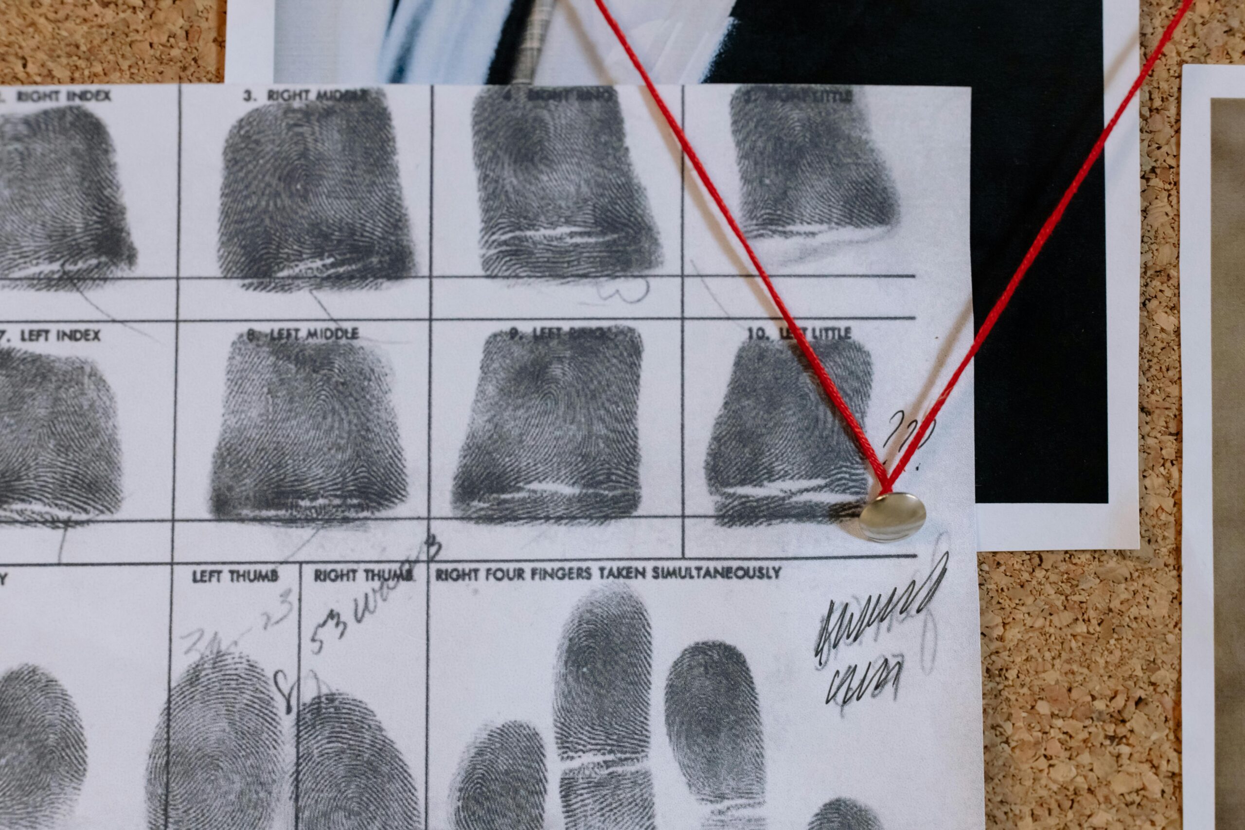
Image-based cell matching represents a revolutionary frontier in biological research, merging advanced computational techniques with microscopy to identify and classify cellular structures with unprecedented precision.
🔬 The Foundation of Image-Based Cell Matching Technology
The convergence of artificial intelligence and cellular biology has created remarkable opportunities for researchers and clinicians alike. Image-based cell matching utilizes sophisticated algorithms to analyze microscopic images, comparing cellular features across vast databases to identify patterns, anomalies, and similarities that would be impossible for human observers to detect consistently.
This technology leverages machine learning models trained on millions of cellular images, enabling automated recognition of cell types, disease markers, and morphological characteristics. The process involves capturing high-resolution images of cells, extracting relevant features, and matching these against reference databases using complex pattern recognition algorithms.
The significance of this approach extends beyond simple identification. It enables researchers to track cellular changes over time, identify rare cell populations within heterogeneous samples, and detect subtle variations that may indicate disease progression or treatment response.
Core Principles Driving Cellular Image Analysis
Understanding the fundamental principles behind image-based cell matching is essential for appreciating both its capabilities and limitations. These principles form the backbone of every successful implementation in laboratory and clinical settings.
Feature Extraction and Pattern Recognition
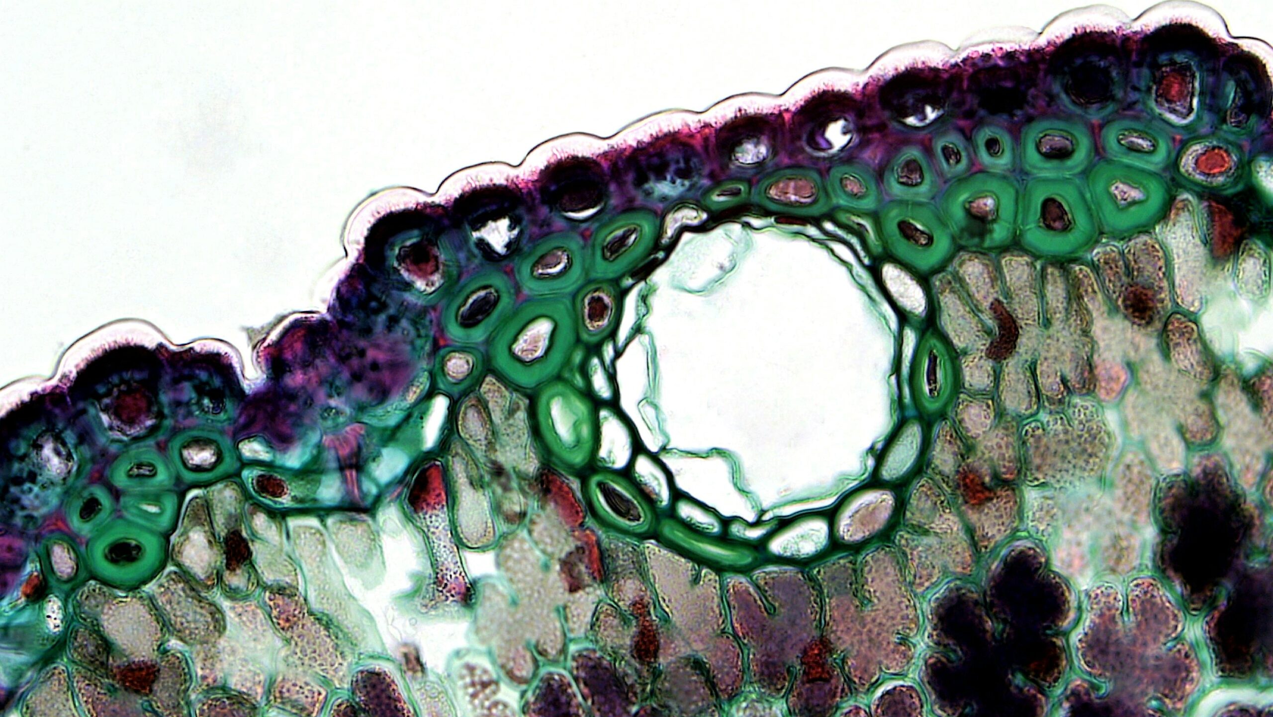
At the heart of cell matching lies the ability to extract meaningful features from microscopic images. These features include cell size, shape, texture, intensity distribution, and spatial relationships with neighboring cells. Advanced algorithms can identify hundreds of distinct features simultaneously, creating comprehensive cellular fingerprints.
Modern deep learning approaches have revolutionized this process by automatically learning which features are most relevant for specific classification tasks. Convolutional neural networks, in particular, excel at recognizing hierarchical patterns in cellular images, from basic edges and textures to complex morphological structures.
Database Architecture and Reference Libraries
The effectiveness of any matching system depends critically on the quality and comprehensiveness of its reference database. These databases must contain accurately labeled examples spanning the full spectrum of cellular variations encountered in real-world applications.
Curating these databases presents significant challenges. Cells exhibit remarkable diversity even within the same type, influenced by factors such as culture conditions, preparation methods, and imaging parameters. Building robust reference libraries requires careful standardization of protocols and extensive validation by expert cytologists.
🎯 Practical Applications Transforming Research and Medicine
The potential applications of image-based cell matching span virtually every domain of biological and medical research. From basic science to clinical diagnostics, this technology is reshaping how we understand and interact with cellular systems.
Cancer Diagnostics and Precision Oncology
In oncology, accurate identification and classification of cancer cells is paramount for diagnosis, prognosis, and treatment selection. Image-based matching systems can analyze tumor biopsies to identify specific cancer subtypes, predict treatment response, and detect minimal residual disease after therapy.
These systems can distinguish between morphologically similar cancer cells based on subtle features that correlate with genetic profiles or drug sensitivity. This capability supports the growing field of precision oncology, where treatment decisions are tailored to the unique characteristics of individual tumors.
Drug Discovery and Toxicology Screening
Pharmaceutical companies increasingly rely on high-content imaging and automated cell matching to screen potential drug candidates. By analyzing how cells respond to different compounds—changes in morphology, protein localization, or organelle structure—researchers can identify promising therapeutic agents and flag potentially toxic substances.
This approach dramatically accelerates the drug development pipeline while reducing costs and animal testing. Automated systems can process thousands of compounds daily, identifying candidates worthy of further investigation based on their cellular effects.
Stem Cell Research and Regenerative Medicine
Stem cell biology presents unique challenges for cell identification and quality control. Image-based matching helps researchers verify stem cell identity, monitor differentiation processes, and ensure the purity of cell preparations intended for therapeutic use.
As regenerative medicine advances toward clinical applications, reliable methods for characterizing stem cell products become essential. Automated image analysis provides objective, reproducible assessments that complement traditional molecular and functional assays.
⚙️ Technical Challenges Requiring Innovative Solutions
Despite impressive advances, image-based cell matching faces significant technical hurdles that limit its broader adoption and effectiveness. Addressing these challenges requires ongoing innovation in both hardware and software domains.
Image Quality and Standardization Issues
Variability in image acquisition represents one of the most persistent challenges. Different microscopes, cameras, lighting conditions, and sample preparation methods all introduce variations that can confound matching algorithms trained on standardized datasets.
Staining techniques pose particular difficulties. The same cell type can appear drastically different depending on whether it’s imaged using brightfield microscopy, phase contrast, fluorescence, or specialized staining protocols. Developing algorithms robust to these variations requires extensive training data and sophisticated normalization techniques.
Computational Demands and Processing Speed
High-resolution microscopy generates enormous datasets. A single experiment might produce hundreds of gigabytes of image data requiring analysis. Processing these volumes demands substantial computational resources, often necessitating specialized hardware like graphics processing units (GPUs) or cloud computing infrastructure.
Real-time analysis presents even greater challenges. Clinical applications, such as intraoperative tumor margin assessment, require results within minutes rather than hours. Balancing accuracy against speed remains an active area of algorithmic development.
Handling Cellular Heterogeneity and Rare Populations
Biological samples rarely contain uniform cell populations. Tissues contain diverse cell types in varying proportions, and disease states often involve rare but clinically significant cell populations. Matching algorithms must reliably identify these rare cells without generating excessive false positives.
Class imbalance—when some cell types are vastly more common than others—creates training difficulties for machine learning models. Specialized techniques like synthetic minority oversampling and cost-sensitive learning help address this issue, but achieving optimal performance across all cell types remains challenging.
📊 Evaluating Performance: Metrics That Matter
Assessing the accuracy and reliability of cell matching systems requires appropriate performance metrics. Different applications prioritize different aspects of system performance, making metric selection crucial.
| Metric | Definition | Primary Use Case |
|---|---|---|
| Accuracy | Overall correct classification rate | Balanced datasets with equal class importance |
| Sensitivity | True positive rate for specific cell type | Detecting rare disease cells |
| Specificity | True negative rate | Minimizing false alarms |
| F1 Score | Harmonic mean of precision and recall | Imbalanced datasets |
| AUC-ROC | Area under receiver operating curve | Threshold-independent evaluation |
Beyond numerical metrics, clinical validation requires demonstrating that automated systems match or exceed human expert performance in real-world conditions. This often involves blinded studies where both algorithms and pathologists analyze identical samples, with discordant cases adjudicated by consensus review.
🚀 Emerging Trends Shaping the Future
The field of image-based cell matching continues to evolve rapidly, driven by advances in artificial intelligence, imaging technology, and biological understanding. Several emerging trends promise to expand capabilities and applications significantly.
Multi-Modal Integration and Data Fusion
Modern approaches increasingly combine imaging data with complementary information sources. Integrating microscopy with genomic profiling, flow cytometry, or clinical metadata enables richer characterizations than imaging alone can provide.
These multi-modal systems learn relationships between visual features and molecular characteristics, predicting genetic profiles or protein expression patterns directly from cellular images. This capability could eventually reduce the need for expensive molecular testing in some clinical scenarios.
Explainable AI and Interpretability
As machine learning models grow more complex, understanding why they make specific classifications becomes increasingly difficult. This “black box” problem concerns clinicians who require explanations for diagnostic decisions that affect patient care.
Explainable AI techniques address this issue by highlighting which image regions most influenced classification decisions or generating human-readable justifications. These approaches build trust in automated systems while potentially revealing new biological insights about cellular characteristics associated with specific conditions.
Federated Learning and Privacy-Preserving Collaboration
Training robust models requires diverse datasets from multiple institutions, but sharing patient data raises privacy concerns. Federated learning enables collaborative model training without centralizing sensitive data—algorithms travel to the data rather than vice versa.
This approach allows hospitals and research centers worldwide to contribute to model improvement while maintaining data security and regulatory compliance. As federated learning matures, it promises to democratize access to state-of-the-art cell matching capabilities.
💡 Best Practices for Implementation Success
Organizations seeking to implement image-based cell matching systems can maximize success probability by following established best practices informed by early adopters’ experiences.
Starting with Clear Objectives and Use Cases
Successful implementations begin with well-defined problems and realistic expectations. Rather than attempting to solve every possible classification challenge simultaneously, focusing on specific high-value use cases allows teams to develop expertise and demonstrate value incrementally.
Engaging end users—pathologists, researchers, or clinicians—from project inception ensures the system addresses genuine needs and integrates smoothly into existing workflows. User feedback during development prevents costly redesigns after deployment.
Investing in Data Quality and Annotation
The adage “garbage in, garbage out” applies forcefully to machine learning systems. High-quality training data with accurate expert annotations forms the foundation of effective models. Organizations should allocate sufficient resources for data curation, recognizing it as an ongoing process rather than a one-time effort.
Establishing standardized imaging protocols minimizes technical variability that could confound analysis. Documentation of acquisition parameters, sample preparation methods, and quality control procedures ensures reproducibility and facilitates troubleshooting when performance issues arise.
Continuous Monitoring and Model Updating
Deploying a cell matching system is not the endpoint but the beginning of an iterative improvement process. Performance should be continuously monitored, with particular attention to cases where the system performs poorly or where human experts disagree with automated classifications.
As new cell types, disease variants, or imaging modalities emerge, models require updating to maintain relevance. Establishing processes for periodic retraining with expanded datasets ensures systems evolve alongside advancing biological knowledge and clinical practice.
🌐 Overcoming Adoption Barriers in Clinical Settings
Despite compelling technical capabilities, translating image-based cell matching from research laboratories to routine clinical use faces substantial non-technical obstacles. Understanding and addressing these barriers is essential for realizing the technology’s full potential.
Regulatory Approval and Validation Requirements
Medical devices incorporating AI algorithms face rigorous regulatory scrutiny. Demonstrating safety and effectiveness through clinical trials requires significant investment and expertise. The evolving regulatory landscape for AI-based diagnostics adds uncertainty, as guidance continues developing in parallel with the technology.
Validation studies must demonstrate consistent performance across diverse patient populations, imaging equipment, and clinical settings. This geographic and demographic generalizability is crucial but challenging to achieve, particularly for rare diseases where large validation cohorts are difficult to assemble.
Workflow Integration and User Acceptance
Introducing automated systems into established clinical workflows requires careful change management. Users need training not only in system operation but in understanding its strengths, limitations, and appropriate use cases. Building confidence that automation enhances rather than replaces human expertise helps overcome resistance.
Interface design profoundly affects acceptance. Systems requiring extensive manual preprocessing or producing outputs in unfamiliar formats create friction that discourages adoption. Seamless integration with existing laboratory information systems and intuitive visualization of results facilitate acceptance.
Looking Ahead: The Transformative Promise
Image-based cell matching stands poised to fundamentally transform how we diagnose disease, develop therapies, and understand cellular biology. As algorithms grow more sophisticated, imaging technology improves, and datasets expand, system capabilities will continue advancing.
The democratization of these tools represents perhaps the most exciting prospect. Cloud-based platforms and user-friendly interfaces are making advanced cell analysis accessible to researchers and clinicians lacking specialized computational expertise. This accessibility could accelerate discoveries and improve patient care globally, particularly in resource-limited settings where expert pathologists are scarce.
Realizing this vision requires continued collaboration across disciplines—computer scientists, biologists, clinicians, ethicists, and regulatory experts must work together to develop systems that are not only technically capable but also safe, fair, and aligned with human values. The journey is challenging, but the potential rewards for science and medicine are immense.
By navigating both the technical principles and practical challenges thoughtfully, the scientific community can unlock the full potential of image-based cell matching, ushering in a new era of precision biology and medicine that benefits researchers, clinicians, and ultimately patients worldwide.