Microscopy has revolutionized biological and materials science, yet the most valuable information often remains hidden within complex image data, waiting to be extracted.
Modern microscopy generates vast amounts of high-resolution image data, but extracting meaningful features from these images presents significant challenges. Traditional analysis methods often fall short when dealing with subtle patterns, three-dimensional structures, or time-lapse sequences. Advanced feature extraction techniques have emerged as powerful tools to unlock this invisible information, enabling researchers to quantify cellular behaviors, identify disease markers, and understand material properties at unprecedented scales.
The field of microscopy image analysis has evolved dramatically over the past decade, driven by advances in computational power, machine learning algorithms, and sophisticated imaging hardware. Scientists can now detect features that were previously imperceptible to human observers, transforming qualitative observations into quantitative data that drives discovery across multiple disciplines.
🔬 The Foundation: Understanding Feature Extraction in Microscopy
Feature extraction refers to the process of identifying and quantifying specific characteristics within microscopy images that provide biological or physical relevance. Unlike simple image measurements like brightness or contrast, advanced features capture complex relationships between pixels, spatial patterns, and temporal dynamics that reveal underlying mechanisms.
The process begins with preprocessing steps that enhance image quality and remove artifacts. This includes background correction, noise reduction, and illumination normalization—essential steps that ensure subsequent analysis produces reliable results. Without proper preprocessing, even the most sophisticated algorithms can produce misleading conclusions.
Modern feature extraction operates at multiple levels simultaneously. Low-level features include edges, corners, and textures that describe basic image properties. Mid-level features capture shapes, regions, and spatial relationships between objects. High-level features encode semantic information about cell types, tissue organization, or material phases that require contextual understanding.
The Challenge of Scale and Dimensionality
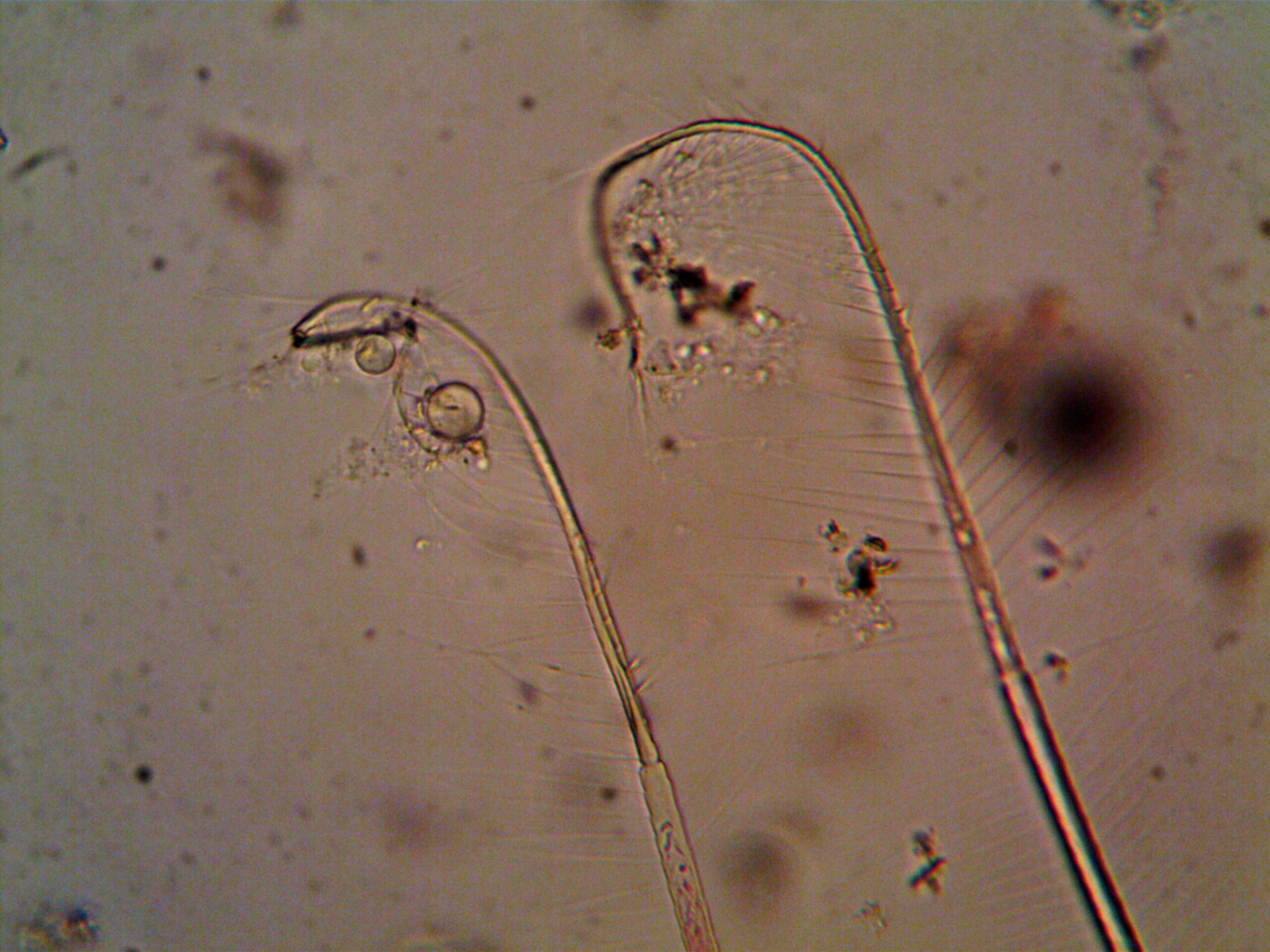
Microscopy images present unique challenges due to their multi-scale nature. Cellular structures span orders of magnitude, from nanometer-scale protein complexes to millimeter-scale tissue architectures. Effective feature extraction must operate across these scales, identifying relevant patterns regardless of their size or position within the image.
Additionally, modern microscopy often generates multi-dimensional datasets including spatial coordinates (x, y, z), time, spectral channels, and even molecular specificity. Extracting features from these high-dimensional spaces requires specialized techniques that can handle computational complexity while preserving biological relevance.
🧮 Classical Computer Vision Approaches
Before the deep learning revolution, classical computer vision techniques formed the backbone of microscopy image analysis. These methods remain relevant today, particularly when data is limited or interpretability is paramount.
Texture analysis through methods like Gray-Level Co-occurrence Matrices (GLCM) quantifies spatial patterns in pixel intensities. These features capture information about homogeneity, contrast, and directionality that often correlate with biological states. For example, nuclear texture features can distinguish between healthy and cancerous cells based on chromatin organization patterns.
Morphological features describe the shape and structure of segmented objects. Parameters like area, perimeter, circularity, and eccentricity provide quantitative descriptions of cellular and subcellular components. Advanced morphological analysis includes skeleton analysis for filamentous structures and convex hull properties for characterizing irregular boundaries.
Intensity-Based Feature Descriptors
Intensity distribution within regions of interest reveals critical biological information. Histogram-based features capture the overall distribution of pixel values, while moment-based features describe asymmetry and peakedness of intensity patterns. Granulometry measures the size distribution of bright structures, useful for characterizing punctate organelles or fluorescent spots.
Gradient-based features detect edges and directional changes in intensity. The Scale-Invariant Feature Transform (SIFT) and Speeded-Up Robust Features (SURF) identify distinctive points that remain recognizable across different imaging conditions, enabling image registration and tracking applications.
🤖 Machine Learning Revolutionizes Feature Discovery
The integration of machine learning into microscopy analysis has fundamentally changed how researchers approach feature extraction. Rather than manually designing features based on domain knowledge, algorithms can automatically discover relevant patterns directly from data.
Convolutional Neural Networks (CNNs) have become the gold standard for extracting features from microscopy images. These architectures learn hierarchical representations, with early layers detecting simple edges and textures, while deeper layers recognize complex patterns specific to the training data. Pre-trained networks like ResNet, VGG, and Inception can be adapted to microscopy applications through transfer learning, reducing the need for large annotated datasets.
The intermediate layers of trained CNNs serve as powerful feature extractors. By extracting activations from specific layers, researchers obtain high-dimensional feature vectors that encode rich information about image content. These features often outperform hand-crafted descriptors, especially for complex classification and segmentation tasks.
Unsupervised Learning for Pattern Discovery
When labeled data is scarce or biological ground truth is unknown, unsupervised learning techniques excel at discovering hidden structure within microscopy datasets. Autoencoders compress images into low-dimensional latent representations, capturing essential features while discarding noise and irrelevant variation.
Variational autoencoders (VAEs) and their variants provide probabilistic frameworks for feature learning, enabling generation of synthetic microscopy images and interpolation between different cellular states. These models reveal continuous spectra of phenotypes rather than discrete categories, better reflecting biological reality.
Clustering algorithms applied to extracted features identify distinct subpopulations within heterogeneous samples. This approach has proven valuable for single-cell analysis, where subtle differences in morphology or intensity patterns distinguish cell types or functional states without prior labeling.
📊 Spatial Statistics and Neighborhood Analysis
Biology occurs in spatial contexts where the arrangement and proximity of structures carries functional significance. Spatial feature extraction captures these relationships through various statistical approaches.
Point pattern analysis quantifies the distribution of objects within images, distinguishing between random, clustered, and regular arrangements. Ripley’s K-function and pair correlation functions measure spatial relationships at multiple scales, revealing organizational principles of subcellular structures or tissue architecture.
Neighborhood features characterize each object based on its local environment. The number, distance, and types of neighboring structures provide contextual information that enhances classification accuracy. For instance, a cell’s behavior often depends not just on its intrinsic properties but also on surrounding cells and extracellular matrix components.
Graph-Based Representations
Representing microscopy images as graphs offers powerful abstraction for feature extraction. Nodes represent individual objects (cells, nuclei, organelles), while edges encode spatial relationships or interactions. Graph-theoretic measures like degree distribution, clustering coefficient, and centrality capture organizational patterns at tissue and cellular scales.
Graph neural networks extend deep learning to graph-structured data, enabling end-to-end learning of spatial features. These architectures naturally incorporate neighborhood information and have shown promise for applications like cell tracking, lineage analysis, and tissue phenotyping.
⏱️ Temporal Features for Dynamic Microscopy
Time-lapse microscopy reveals cellular processes as they unfold, but extracting meaningful features from temporal sequences requires specialized techniques. Motion patterns, growth rates, and dynamic responses to perturbations provide insights impossible to obtain from static images.
Optical flow algorithms track movement of intensity patterns between frames, quantifying velocity fields that describe cellular migration, cytoplasmic streaming, or organelle transport. These flow features characterize both individual object trajectories and collective behaviors in populations.
Temporal texture analysis extends spatial texture methods to include time as an additional dimension. Local Binary Patterns applied to space-time volumes detect dynamic patterns like oscillations, waves, or transient events that distinguish different biological processes.
Tracking and Trajectory Analysis
Linking detected objects across time frames enables trajectory-based feature extraction. Track features include displacement, velocity, directionality, and confinement ratio that characterize movement patterns. Higher-order analysis examines changes in these parameters over time, detecting acceleration, turning behavior, or transitions between movement modes.
Hidden Markov Models and change-point detection identify distinct behavioral states within trajectories. A cell might alternate between exploratory and confined movements, with transition frequencies and state durations serving as phenotypic features that respond to experimental manipulations.
🎨 Multi-Channel and Spectral Feature Extraction
Fluorescence microscopy often employs multiple channels to simultaneously visualize different molecular species. Extracting features that capture relationships between channels reveals colocalization, stoichiometry, and functional interactions.
Colocalization analysis quantifies spatial overlap between fluorescent signals. Beyond simple overlap coefficients, advanced methods consider intensity correlations, object-based colocalization, and statistical significance testing to distinguish true biological associations from random overlap.
Spectral unmixing separates overlapping emission spectra into constituent components, enabling quantification of multiple markers even when their signals overlap. Features extracted from unmixed channels provide more accurate quantification than raw multi-channel images, particularly in densely labeled samples.
Radiomics Applied to Microscopy
Radiomics, originally developed for medical imaging analysis, extracts large numbers of quantitative features from images. Applied to microscopy, radiomic approaches compute hundreds of features spanning intensity, texture, shape, and wavelet domains, creating comprehensive phenotypic profiles.
Feature selection methods then identify the subset of radiomic features most relevant for specific biological questions. This high-throughput phenotyping approach has proven effective for drug screening, disease classification, and identifying biomarkers in complex microscopy datasets.
🔍 Deep Feature Extraction Beyond Standard CNNs
While standard CNNs excel at many tasks, specialized architectures address unique challenges in microscopy image analysis. U-Net and its variants perform semantic segmentation while preserving spatial resolution, extracting features that maintain precise localization necessary for quantitative analysis.
Attention mechanisms highlight image regions most relevant for classification or other tasks, providing both improved performance and interpretability. Spatial attention maps reveal which cellular structures or tissue regions drive predictions, offering biological insights alongside quantitative features.
Self-supervised learning trains feature extractors without manual annotations by solving pretext tasks like image rotation prediction, context encoding, or contrastive learning. These approaches leverage vast amounts of unlabeled microscopy data to learn representations that transfer effectively to downstream analysis tasks.
Foundation Models for Microscopy
Large-scale foundation models trained on diverse microscopy datasets are emerging as universal feature extractors. Similar to how BERT transformed natural language processing, these models learn general-purpose representations that capture fundamental properties of cellular and tissue organization across different imaging modalities and biological systems.
Fine-tuning foundation models for specific applications requires minimal labeled data while achieving state-of-the-art performance. This democratizes advanced microscopy analysis, making sophisticated feature extraction accessible to laboratories without extensive machine learning expertise or computational resources.
💡 Practical Considerations and Best Practices
Implementing advanced feature extraction requires careful attention to experimental design and computational workflow. Image quality remains paramount—no algorithm can extract meaningful features from poorly acquired images. Proper calibration, optimal exposure settings, and appropriate sampling rates in time-lapse experiments ensure data quality sufficient for quantitative analysis.
Feature standardization and normalization prevent technical variation from overwhelming biological signal. Batch effects from different imaging sessions or instruments can introduce spurious patterns that obscure true biological differences. Appropriate normalization strategies and batch correction methods ensure extracted features reflect biology rather than technical artifacts.
Validation through independent datasets and orthogonal methods establishes confidence in extracted features. Cross-validation prevents overfitting, while comparison with manual annotations or alternative computational approaches confirms biological relevance. Negative controls and positive controls help distinguish signal from noise.
Computational Infrastructure and Scalability
Modern microscopy generates data at rates that challenge traditional analysis pipelines. Cloud computing and GPU acceleration enable processing of large datasets, while containerization ensures reproducibility across different computing environments. Open-source frameworks like ImageJ, CellProfiler, and Python-based libraries provide accessible tools for implementing advanced feature extraction.
Automated pipelines that integrate acquisition, processing, and analysis reduce manual intervention and improve throughput. Real-time feature extraction during acquisition enables adaptive microscopy experiments where imaging parameters adjust based on extracted features, optimizing data collection for specific biological questions.
🌟 Emerging Frontiers and Future Directions
The field continues rapid evolution with several exciting developments on the horizon. Physics-informed neural networks incorporate optical principles and biological constraints into feature learning, improving generalization and interpretability. These hybrid approaches combine data-driven learning with domain knowledge, potentially overcoming limitations of purely empirical methods.
Multimodal integration combines microscopy with other measurement modalities like genomics, proteomics, or metabolomics. Features extracted across modalities provide holistic cellular and tissue characterization, revealing relationships between molecular composition, spatial organization, and function that single-modality approaches miss.
Explainable AI techniques address the black-box nature of deep learning feature extractors. Methods like layer-wise relevance propagation and concept activation vectors decompose complex features into interpretable components, building trust and enabling biological insight from learned representations.
As microscopy technology advances with higher resolution, faster acquisition, and expanded molecular specificity, feature extraction methods must evolve in parallel. The integration of advanced computational techniques with cutting-edge imaging promises to unlock ever more invisible information, transforming our understanding of biological systems at every scale. The future of microscopy analysis lies not just in seeing more clearly, but in extracting meaning from visual complexity through intelligent computational approaches that reveal the hidden patterns governing life itself.