Microscopy has revolutionized botanical forensics, transforming tiny plant fragments into powerful evidence that solves crimes and uncovers environmental mysteries through cellular-level investigation.
🔬 The Invisible World of Plant Evidence
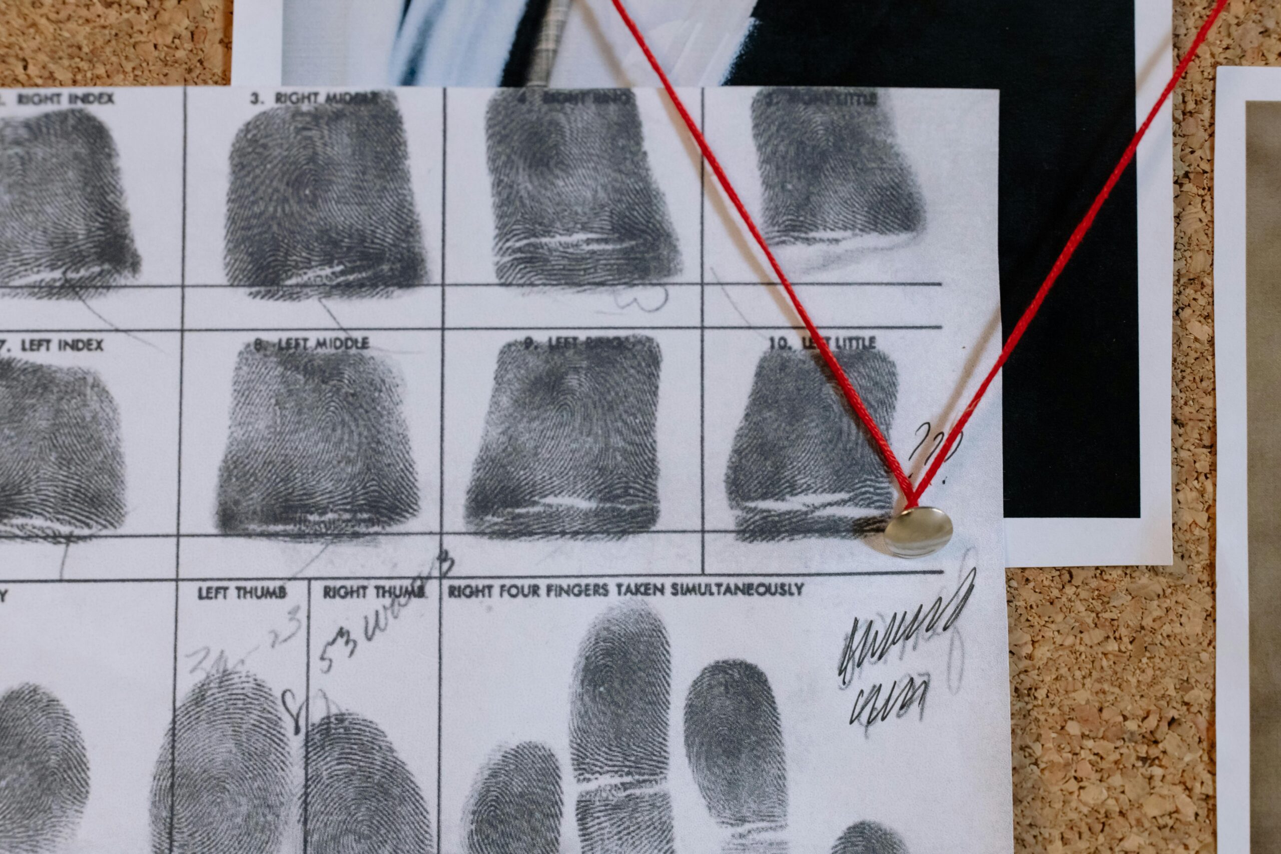
When investigators arrive at a crime scene, they’re not just looking for fingerprints or DNA evidence. Hidden in plain sight are botanical clues that can place a suspect at a specific location, determine time of death, or even reconstruct entire criminal timelines. These microscopic plant materials—pollen grains, spores, wood fragments, and cellular structures—tell stories that are invisible to the naked eye but crystal clear under the lens of a microscope.
Botanical forensics, also known as forensic botany, represents the intersection of plant science and criminal investigation. This specialized field has gained prominence in recent decades as microscopy technology has advanced, allowing forensic botanists to examine plant evidence with unprecedented detail and accuracy. From solving murders to tracking illegal timber trade, microscopic plant analysis has become an indispensable tool in modern forensic science.
Understanding the Fundamentals of Forensic Botany
Forensic botany encompasses the application of plant science to legal investigations. Every environment contains unique botanical signatures—specific combinations of plant species, growth patterns, and microscopic structures that can identify locations with remarkable precision. When a person or object comes into contact with vegetation, microscopic plant materials transfer and adhere to clothing, shoes, vehicles, and other surfaces.
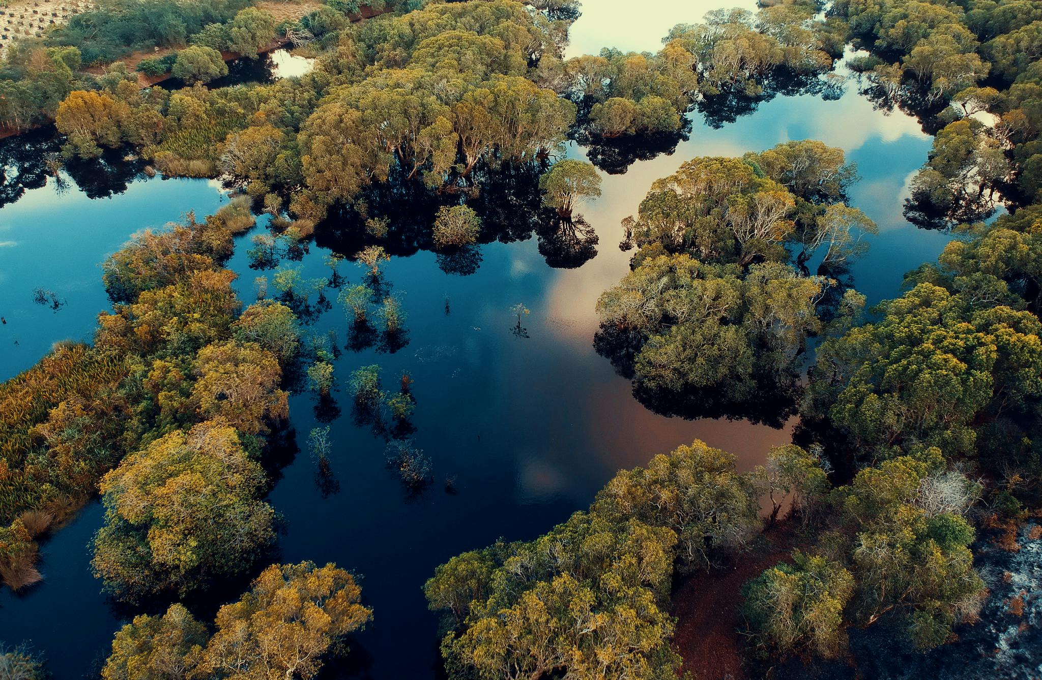
These microscopic transfers create a botanical fingerprint. Pollen grains, which measure between 10 and 100 micrometers, are particularly valuable because they’re extremely durable, species-specific, and ubiquitous in the environment. A single square centimeter of soil can contain thousands of pollen grains representing dozens of plant species, creating a detailed botanical profile of a specific location.
The Power of Microscopic Plant Structures
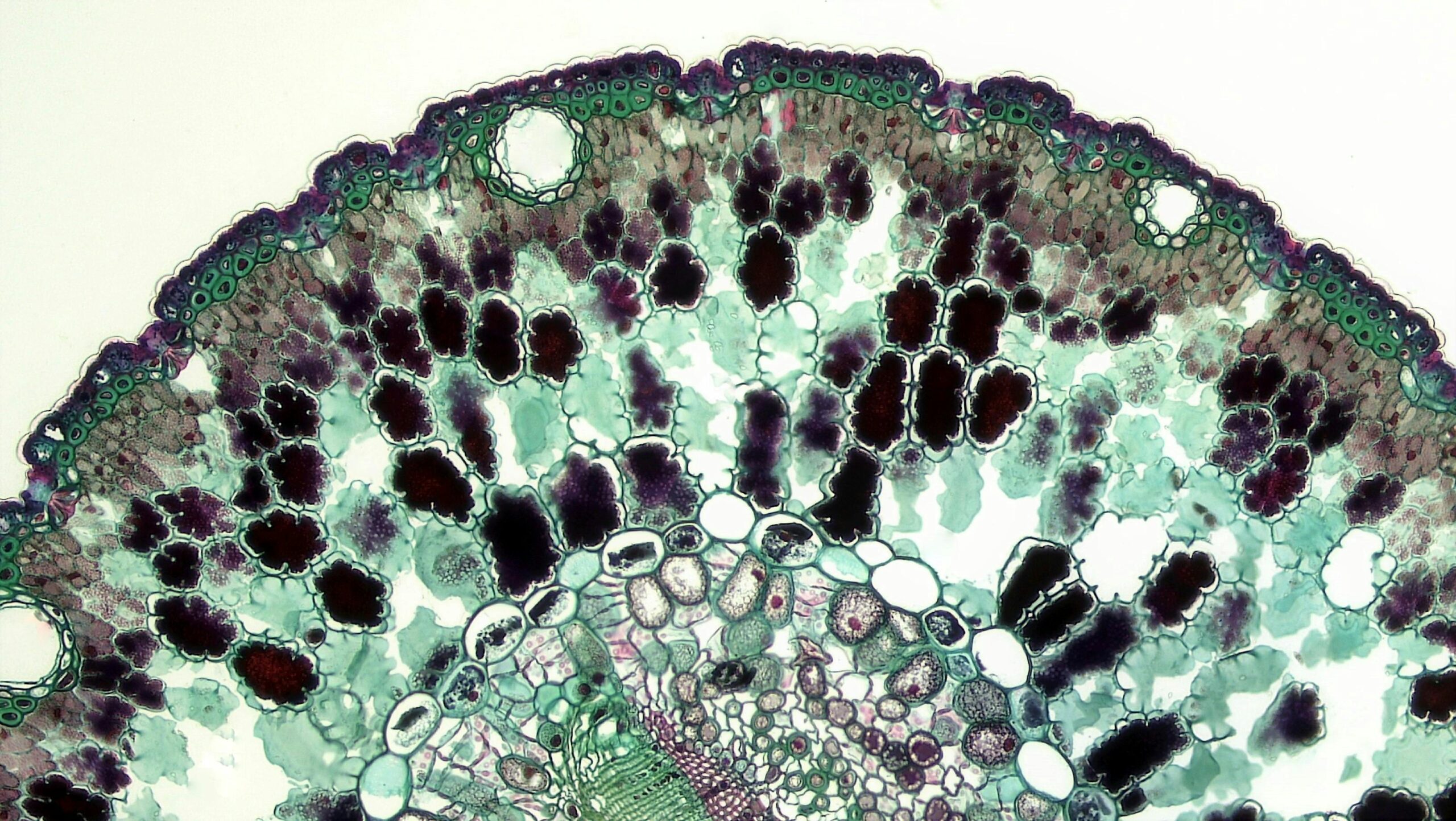
Plant cells possess distinctive features that make them invaluable for forensic identification. Cell wall patterns, chloroplast arrangements, trichome structures, and vascular tissue organization vary dramatically between species. Under microscopic examination, these cellular characteristics become diagnostic tools that can identify plant species, sometimes down to individual varieties.
Wood anatomy provides particularly robust evidence. The arrangement of vessels, fibers, and ray cells creates unique patterns that forensic botanists can use to identify tree species and sometimes even determine geographic origin. This capability has proven essential in combating illegal logging and timber fraud, where microscopic wood analysis can verify whether expensive hardwood truly originated from claimed sustainable sources.
🌿 Types of Microscopy Techniques in Botanical Forensics
Modern botanical forensics employs multiple microscopy techniques, each offering unique advantages for examining different types of plant evidence. The selection of appropriate microscopy methods depends on the nature of the evidence, the questions being investigated, and the level of detail required.
Light Microscopy and Compound Microscopes
Traditional light microscopy remains the workhorse of forensic botany. Compound microscopes use visible light and multiple lenses to magnify specimens up to 1000 times, revealing cellular structures, pollen morphology, and tissue organization. Polarized light microscopy adds another dimension by exploiting the birefringent properties of plant structures, particularly useful for identifying starch grains and cellulose fibers.
Phase contrast microscopy enhances the visibility of transparent specimens without staining, making it ideal for examining fresh plant tissues and living cells. Differential interference contrast (DIC) microscopy creates three-dimensional-appearing images that highlight surface features and internal structures with exceptional clarity.
Electron Microscopy for Ultimate Resolution
When greater magnification and resolution are needed, forensic botanists turn to electron microscopy. Scanning electron microscopy (SEM) provides detailed three-dimensional images of surface structures at magnifications exceeding 100,000 times. SEM reveals the intricate architecture of pollen grain surfaces, trichome structures, and epidermal cell patterns that are diagnostic for species identification.
Transmission electron microscopy (TEM) penetrates through specimens, revealing internal cellular ultrastructure including organelle organization and cell wall composition. While less commonly used in routine forensic work due to complex sample preparation requirements, TEM provides unmatched detail for specialized investigations.
Fluorescence Microscopy and Chemical Analysis
Fluorescence microscopy exploits the natural autofluorescence of plant compounds or uses fluorescent dyes to highlight specific cellular components. Lignin, chlorophyll, and various secondary metabolites exhibit characteristic fluorescence patterns that aid in plant identification and tissue analysis.
Modern fluorescence techniques can be combined with spectroscopic analysis to create chemical maps of plant tissues, identifying specific compounds distributed within cellular structures. This capability proves particularly valuable when investigating poisoning cases involving toxic plants or determining the authenticity of herbal products.
📊 Real-World Applications in Criminal Investigations
The practical application of microscopic botanical forensics has solved numerous criminal cases worldwide. These investigations demonstrate how microscopic plant evidence provides objective, scientifically robust information that can corroborate or contradict witness testimony and suspect statements.
Placing Suspects at Crime Scenes
One of the most powerful applications of forensic botany involves linking suspects to specific locations through pollen and seed analysis. When investigators collect dust from a suspect’s vehicle, clothing, or shoes, microscopic examination often reveals a distinctive pollen assemblage. By comparing this botanical profile to pollen samples from crime scenes and alibi locations, forensic botanists can determine whether the suspect visited the crime scene.
In several notable cases, pollen evidence has proven decisive. Unique combinations of local plant species create botanical signatures that can be as location-specific as GPS coordinates. Urban environments, despite appearing botanically homogeneous, often contain distinctive plant communities in parks, gardens, and landscaped areas that create identifiable microscopic signatures.
Determining Time Since Death
Botanical evidence also contributes to estimating postmortem intervals. Plant growth patterns, seed germination, and root penetration through clothing or remains provide temporal information. Microscopic examination of plant materials found with remains can identify seasonal species that bloom or produce pollen during specific times of year, narrowing the window when death occurred.
Succession patterns of algae, fungi, and other microorganisms on submerged remains follow predictable sequences that microscopy can document and analyze. These botanical clocks, while requiring careful interpretation considering environmental variables, complement traditional forensic methods for determining time since death.
🌍 Environmental Crimes and Wildlife Protection
Beyond traditional criminal investigations, microscopic botanical forensics plays an increasingly important role in environmental protection and wildlife crime prevention. The illegal trade in endangered plant species, timber fraud, and contamination cases all benefit from microscopic plant analysis.
Combating Illegal Timber Trade
The global illegal timber trade represents a multi-billion dollar criminal enterprise that devastates forests, undermines legitimate businesses, and funds organized crime. Microscopic wood anatomy provides a scientific tool for verifying timber species and geographic origin. Wood identification requires examining cellular structures in three dimensions—transverse, radial, and tangential sections—each revealing distinctive patterns of vessels, rays, and fibers.
Forensic wood anatomists maintain extensive reference collections and databases documenting the microscopic characteristics of thousands of tree species. When suspicious timber is intercepted, microscopic analysis can determine whether expensive tropical hardwood claimed as sustainably harvested actually came from protected forests, or whether substituted species were fraudulently labeled as more valuable woods.
Protecting Endangered Plant Species
International treaties like CITES (Convention on International Trade in Endangered Species) regulate trade in thousands of plant species. Enforcement requires accurate identification, often from processed products where macroscopic features are no longer visible. Microscopic analysis of wood, fibers, and cellular structures enables identification of endangered species in products ranging from furniture to traditional medicines.
The illegal trade in succulent plants, orchids, and other ornamentals threatens many species with extinction. Microscopic examination of epidermal tissues, trichomes, and vascular structures can identify species even from small fragments, supporting prosecution of wildlife trafficking cases.
🔍 Methodologies and Best Practices
Conducting microscopic botanical forensics requires rigorous protocols to ensure reliability and admissibility in legal proceedings. Chain of custody documentation, contamination prevention, and standardized analytical methods are essential for producing defensible scientific evidence.
Evidence Collection and Preservation
Proper collection of botanical evidence begins at the crime scene. Investigators use specialized techniques to recover pollen and plant fragments without contamination. Sticky tape lifts, vacuum collection with specialized filters, and careful packaging in clean containers prevent cross-contamination and preserve microscopic materials.
Reference samples from the crime scene and surrounding areas provide comparison materials essential for interpretation. Forensic botanists collect soil cores, surface debris, and vegetation samples that document the local botanical community. These reference collections create baseline data against which suspect-associated materials can be compared.
Sample Preparation Techniques
Microscopic examination requires appropriate sample preparation that varies depending on the material and microscopy technique. Pollen extraction uses chemical treatments to remove organic matter and minerals, concentrating pollen grains for easier examination. Acetolysis, a standard technique, treats samples with acetic anhydride and sulfuric acid, dissolving cellular contents while preserving pollen walls.
Wood samples require precise sectioning to reveal diagnostic anatomical features. Microtomes cut sections 10-20 micrometers thick that are then stained and mounted on glass slides. Different staining protocols highlight specific cellular components—safranin stains lignified tissues red while fast green counterstains cellulose walls.
💡 Interpreting Microscopic Plant Evidence
The scientific value of microscopic botanical evidence depends on proper interpretation that considers both the power and limitations of the techniques. Forensic botanists must understand plant biology, ecology, and taxonomy to draw valid conclusions from microscopic observations.
Statistical Analysis and Probability
Modern forensic botany increasingly employs statistical methods to quantify the strength of botanical evidence. Rather than simply noting presence or absence of plant species, forensic botanists analyze pollen assemblages quantitatively, counting hundreds of grains to determine relative abundances of different species.
These quantitative profiles enable statistical comparison using similarity indices and multivariate analysis. Bayesian approaches calculate likelihood ratios—the probability of observing the evidence if the suspect visited the crime scene versus if they did not. This statistical framework provides courts with transparent, objective assessments of evidence strength.
Understanding Limitations and Uncertainties
Honest communication about limitations is crucial for scientific integrity in forensic testimony. Botanical evidence rarely provides absolute certainty but rather narrows possibilities and supports or contradicts hypotheses. Factors like pollen dispersal distances, seasonal variation, and secondary transfer create uncertainties that require careful consideration.
Forensic botanists must also recognize the current limits of taxonomic knowledge. While pollen of many species is distinctively identifiable microscopically, some plant families produce morphologically similar pollen that cannot be reliably distinguished. Reference collections may lack representatives of rare species or regional variants, limiting identification capabilities.
🎓 Training and Expertise Requirements
Conducting microscopic botanical forensics requires extensive training that combines plant taxonomy, microscopy skills, and forensic science principles. Practitioners typically hold advanced degrees in botany, plant biology, or related fields, supplemented by specialized forensic training.
Proficiency in microscopic plant identification demands years of practice examining thousands of specimens. Building mental libraries of diagnostic features—pollen aperture patterns, cell wall thicknesses, trichome morphologies—requires sustained study using authenticated reference materials. Many forensic botanists maintain personal reference collections containing thousands of prepared slides representing local and exotic plant species.
Professional organizations like the International Association of Forensic Sciences provide forums for knowledge exchange and continuing education. Workshops, conferences, and peer-reviewed publications disseminate new techniques and case studies, advancing the field’s collective expertise.
🚀 Future Directions and Emerging Technologies
Microscopy techniques continue advancing, offering forensic botanists ever more powerful analytical capabilities. Digital imaging and automated analysis promise to enhance both the speed and objectivity of microscopic plant identification.
Automated Image Analysis and Machine Learning
Artificial intelligence and machine learning algorithms are being trained to recognize and classify pollen grains and other plant structures from microscopic images. These systems can rapidly process thousands of images, identifying species and quantifying abundances with consistency that complements human expertise.
Automated systems require extensive training datasets containing validated images of known species. Building these databases represents a major undertaking, but once established, they enable rapid, standardized analysis that reduces subjectivity and increases reproducibility.
Integrating Multiple Analytical Approaches
The future of botanical forensics lies in integrating microscopy with complementary techniques including DNA analysis, chemical profiling, and isotope analysis. DNA barcoding can identify species from minute tissue samples, while stable isotope ratios reveal geographic origins. Combining multiple independent lines of evidence creates more robust conclusions than any single technique alone.
Portable microscopy devices and field-deployable analysis systems are emerging that could enable preliminary botanical assessments at crime scenes. While not replacing laboratory analysis, these tools could guide evidence collection and investigative priorities in real time.
🌟 The Unsung Heroes of Justice
Microscopic botanical forensics represents a specialized but invaluable tool in the modern investigative arsenal. The ability to extract meaningful information from tiny plant fragments that others might overlook demonstrates the power of scientific observation and careful analysis. As microscopy technology advances and forensic botanists refine their methods, the hidden stories contained in pollen grains, wood cells, and plant tissues will continue revealing truths that serve justice.
The field exemplifies how fundamental botanical knowledge applies to practical problems with significant societal impact. Every solved case, prevented environmental crime, and protected endangered species testifies to the value of understanding plant biology at the microscopic level. Nature’s secrets, when revealed through the lens of a microscope, illuminate human activities and hold wrongdoers accountable.
For those intrigued by this intersection of biology and justice, botanical forensics offers a career path combining scientific rigor with real-world impact. The next time you brush past vegetation or track soil into your home, consider the microscopic botanical evidence you might be carrying—evidence that, in different circumstances, could reveal where you’ve been and what you’ve encountered. In the hands of skilled forensic botanists armed with microscopes, even the smallest plant fragments become powerful witnesses that cannot lie.